QBio products are professional-use topical cosmetics. Manufactured in our FDA-registered U.S. facility.

QBio · Pre-Clinical Research

QBio Elixir
In vitro findings in human dermal fibroblasts

A pre-clinical laboratory study conducted in collaboration with the Nano Medicine and Tissue Engineering Laboratory at the University of Texas at Arlington, examining the QBio Elixir formulation in cultured human dermal fibroblasts.

Measured in culture, versus untreated controls

Three assays were performed on adult human dermal fibroblasts (HDFa). In each, the QBio Elixir-exposed cells were compared against untreated cells serving as the negative control.

Cell proliferation

MTS assay · over 4 days

15×

Cell migration

Scratch assay · 12 hours

~2×

Collagen I signal

Immunofluorescence · 2 days

Untreated controlQBio Elixir condition

Cell proliferation

Proliferation assay

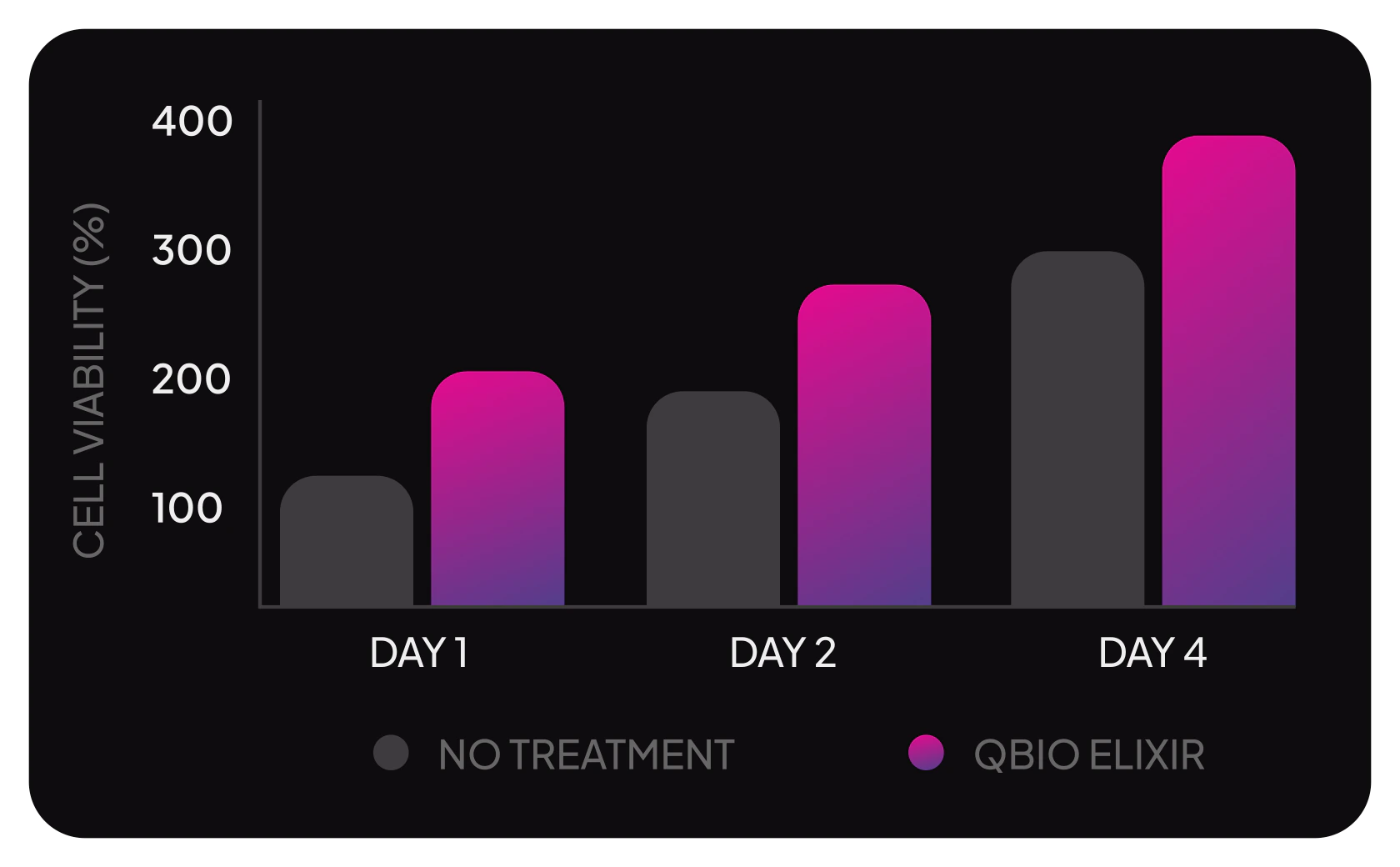
Figure 1. Cell viability (%) at days 1, 2 and 4 in untreated control versus QBio Elixir–exposed human dermal fibroblasts.
Concentration
A dilution of 1:5 was identified as effective for the study.
Observation
Cells in the QBio Elixir condition showed increased growth over several days compared with untreated controls.
Interpretation
The formulation may support cell proliferation in laboratory settings, with the QBio Elixir condition demonstrating up to twice the growth rate of untreated cells.

Cell migration

Scratch migration assay

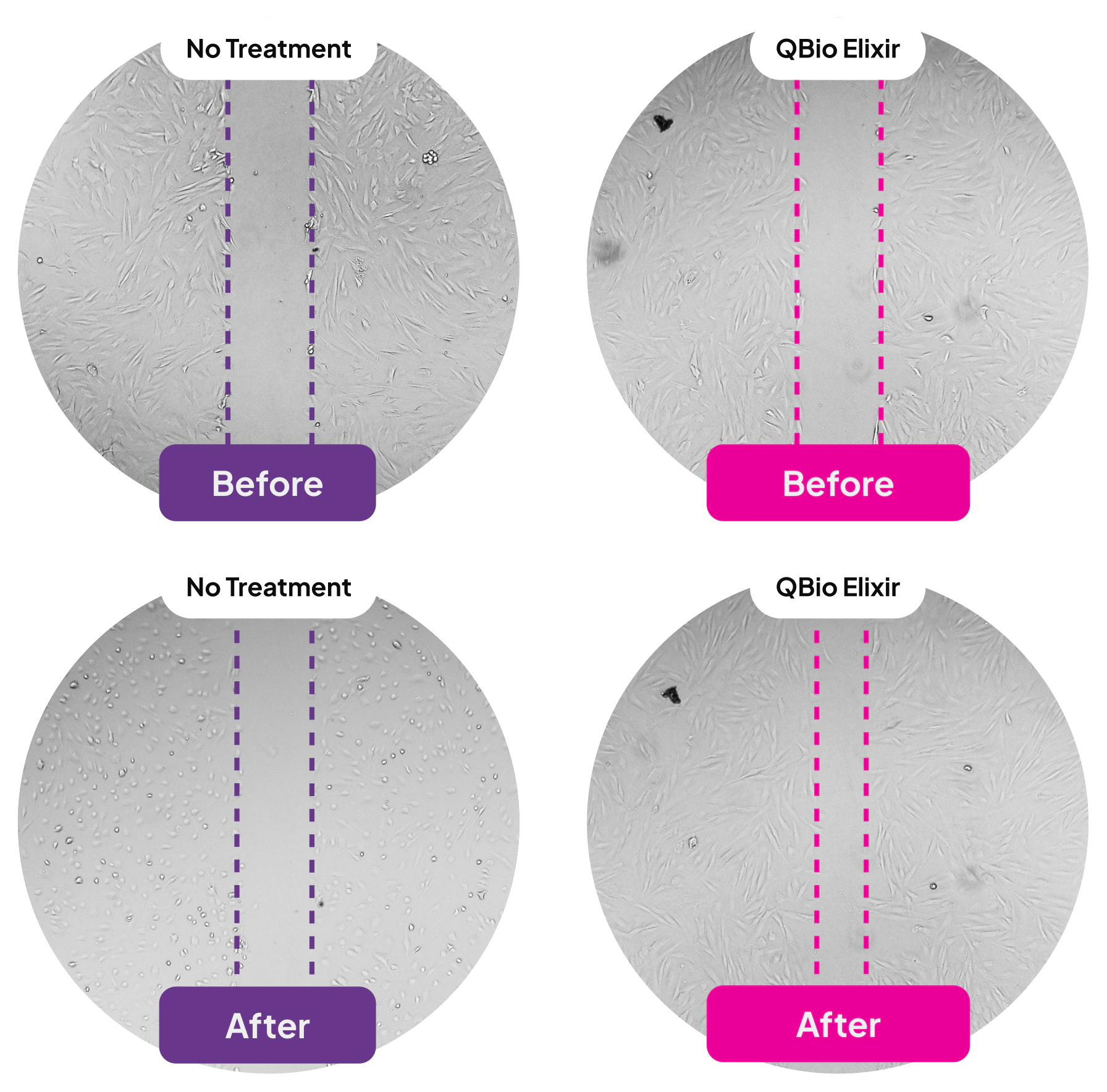
Figure 2. Phase-contrast images of gap closure in a cell-culture scratch assay, untreated control versus QBio Elixir condition, at 0 and 12 hours.
Method
A migration assay compared the QBio Elixir condition with untreated controls.
Observation
Cells in the QBio Elixir condition demonstrated enhanced movement in the assay.
Interpretation
The formulation may promote cell migration in vitro, with the QBio Elixir condition showing up to a 15-fold increase in migration compared with controls.

Collagen I production

Collagen I immunofluorescence

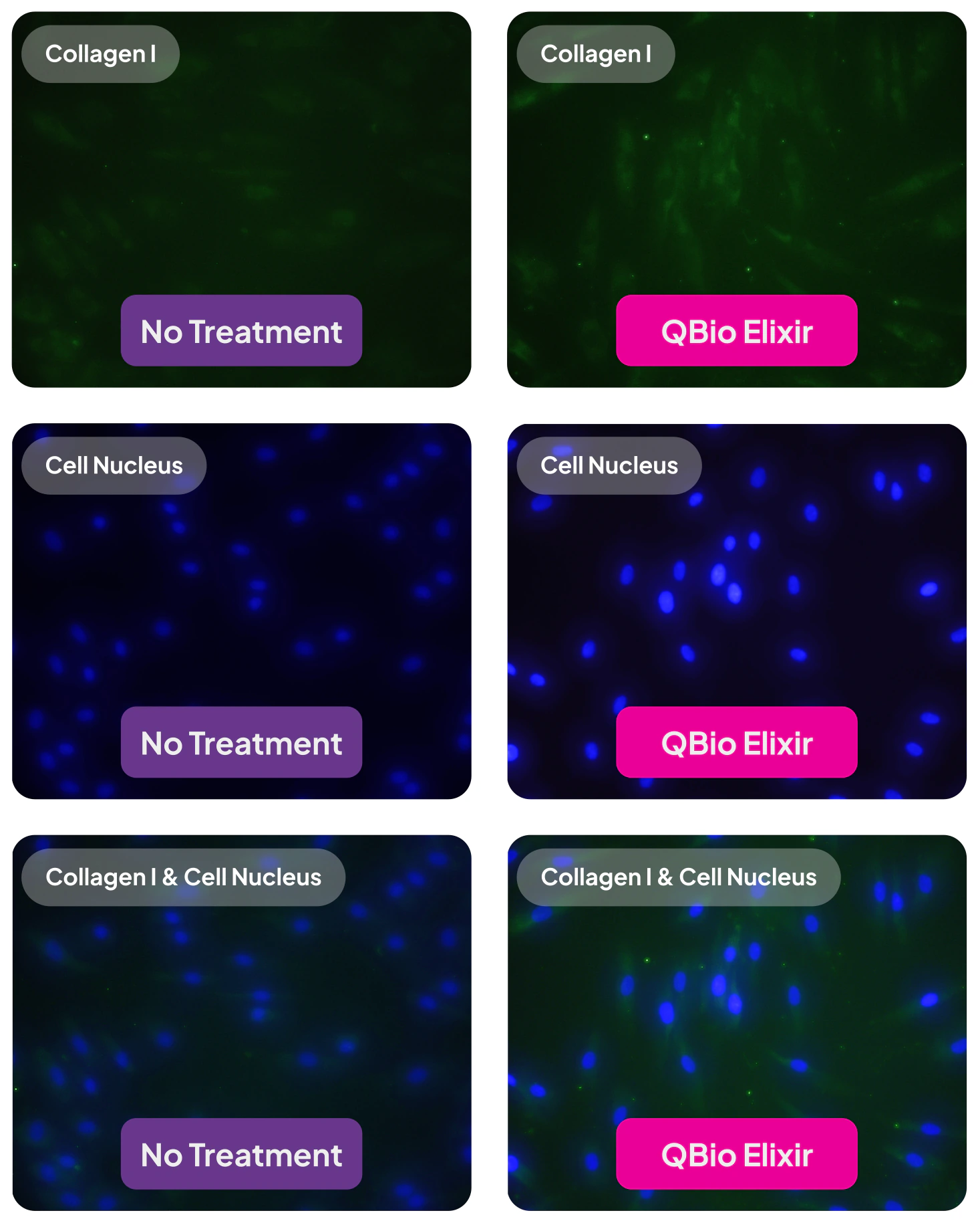
Figure 3. Fluorescent imaging of Collagen I signal and cell nuclei, untreated control versus QBio Elixir–exposed cultures.
Method
Collagen I levels were measured using immunofluorescence techniques.
Observation
An increase in Collagen I signal was observed in the QBio Elixir condition compared with controls.
Interpretation
The formulation may support collagen synthesis in laboratory conditions, with the QBio Elixir condition producing up to twice as much collagen as untreated cells.

Full experimental detail

Complete protocols, cell lines, reagents, and catalog numbers, as reported by the study authors.

Human dermal fibroblast culture

Adult Human Dermal Fibroblasts (HDFa) (ATCC® PCS-201-012), up to passage 10, were cultured in complete Dulbecco's Modified Eagle's Medium (DMEM) supplemented with 10% fetal bovine serum (FBS) and 1% penicillin/streptomycin.

Cells were maintained in a humidified incubator with 95% air and 5% CO₂ at 37 °C. The medium was changed every other day until the cells reached confluence. Sub-culturing was performed when the HDFa culture reached 80% confluency. For in vitro studies and assays, a low-serum medium with 1% FBS was used.

Proliferation study

Cells were seeded at a density of 2,000 cells per well in 96-well plates and incubated overnight. The following day, cells were exposed to QBio Elixir at various concentrations, diluted from 1× to 1:50×, and maintained for up to four days. Media and fresh QBio Elixir were replenished every two days.

Cell growth was assessed using an MTS assay according to the manufacturer's instructions (CellTiter 96® AQueous One Solution Cell Proliferation Assay, Promega, Madison, WI). Untreated cells served as the negative control. Cellular growth was represented as a percentage relative to untreated cells at 24 hours.

Scratch (migration) assay

Reported by the authors under the standard literature term “wound healing (scratch) assay” — a cell-culture migration measurement.

Cells were seeded at a density of 20,000 cells per well in a 48-well plate and incubated overnight. Gaps were created using a 200 µL pipette tip. Cells were washed with phosphate-buffered saline (PBS) and incubated with QBio Elixir at an optimized concentration (1:5 dilution) in low-serum medium for 12 hours at 37 °C.

Phase-contrast images were taken to measure the initial gap distance at 0 hours and the degree of gap closure at 12 hours. Untreated cells served as the negative control. Cell migration was quantified by calculating the average distance traveled relative to the initial gap distance. For each sample in each replicate, six distances were selected and measured to determine migration.

Extracellular matrix (ECM) production

Cells were seeded at a density of 10,000 cells per well on µ-Slide 8-well chambers (80826, ibidi). The next day, cells were exposed to QBio Elixir at a 1:5 dilution for two days, then fixed in cold methanol and permeabilized with 0.1% Triton X-100. After washing with PBS for five minutes three times, cells were blocked at room temperature with 1% bovine serum albumin (BSA) in 0.1% Tween 20 in PBS (TPBS).

Cells were then incubated overnight at 4 °C with primary antibodies: rabbit anti-human Elastin Polyclonal Antibody (1:200, 15257-1-AP, ProteinTech Group), rabbit anti-human Collagen I Antibody (1:200, PA1-26204, Invitrogen), or rabbit anti-human MMP1 Antibody (1:50, 10371-2-AP, ProteinTech Group), diluted in 1% BSA in TPBS.

Following washing, cells were incubated with secondary antibody Goat Anti-Rabbit IgG H&L (FITC) (1:1000, Abcam, ab6717) in 1% BSA in TPBS for one hour at room temperature. Nuclei were counterstained with DAPI for five minutes. Finally, slides were washed, mounted, and imaged using an Echo fluorescent microscope.

Scope and limitations

This work was conducted entirely in vitro, in cultured human dermal fibroblasts. Cell-culture models are a standard early step in cosmetic ingredient science, but they do not replicate intact human skin, and results in culture do not establish outcomes in people.

Observations here describe how cells behaved in laboratory conditions relative to untreated controls. They are not measurements of the finished product's performance on skin, and they are not claims of any therapeutic or medical effect.

The study authors note that further research, including clinical evaluation, would be needed to re-affirm these observations and to understand the formulation's role as part of a cosmetic regimen.

References

  1. Chang, L., Fan, WW., Yuan, HL. et al. Role of umbilical cord mesenchymal stromal cells in skin rejuvenation. npj Regen Med 9, 20 (2024). doi.org/10.1038/s41536-024-00363-1
  2. Fitriani N, Wilar G, Narsa AC, Mohammed AFA, Wathoni N. Application of Amniotic Membrane in Skin Regeneration. Pharmaceutics. 2023;15(3):748. doi: 10.3390/pharmaceutics15030748.
  3. Elkhenany H, El-Derby A, Abd Elkodous M, Salah RA, Lotfy A, El-Badri N. Applications of the amniotic membrane in tissue engineering and regeneration: the hundred-year challenge. Stem Cell Res Ther. 2022;13(1):8.
  4. Vojdani Z, Babaei A, Vasaghi A, Habibagahi M, Talaei-Khozani T. The effect of amniotic membrane extract on umbilical cord blood mesenchymal stem cell expansion. Iran J Basic Med Sci. 2016;19(1):89–96.
  5. Hofmann, N., Rennekampff, H.-O., Salz, A. K., & Börgel, M. Preparation of human amniotic membrane for transplantation in different application areas. Frontiers in Transplantation, 2 (2023). doi.org/10.3389/frtra.2023.1152068
  6. Nejad, A. R., Hamidieh, A. A., Amirkhani, M. A., & Sisakht, M. M. Update review on five top clinical applications of human amniotic membrane in regenerative medicine. Placenta, 103, 104–119 (2021). doi.org/10.1016/j.placenta.2020.10.026
  7. Biswas, A., Rajasekaran, R., Saha, B., et al. Human placenta/umbilical cord derivatives in regenerative medicine – prospects and challenges. Biomaterials Science, 11(14), 4789–4821 (2023). doi.org/10.1039/d2bm01977a
  8. Aratikatla A, Maffulli N, Rodriguez HC, et al. Allogenic perinatal tissue for musculoskeletal regenerative medicine applications: a systematic review. Biomedicines. 2022;10(12):3173.
  9. Cooke M, Tan EK, Mandrycky C, et al. Comparison of cryopreserved amniotic membrane and umbilical cord tissue with dehydrated amniotic membrane/chorion tissue. J Wound Care. 2014;23(10):465–476.
  10. Liu J, Sheha H, Fu Y, Liang L, Tseng SC. Update on amniotic membrane transplantation. Expert Rev Ophthalmol. 2010;5(5):645–661.
  11. Dua HS, Gomes JA, King AJ, Maharajan VS. The amniotic membrane in ophthalmology. Surv Ophthalmol. 2004;49(1):51–77.
  12. Hanselman AE, Tidwell JE, Santrock RD. Cryopreserved human amniotic membrane injection for plantar fasciitis: a randomized, controlled, double-blind pilot study. Foot Ankle Int. 2015;36(2):151–158.
  13. Buck D. Amniotic umbilical cord particulate for discogenic pain. J Osteopath Med. 2019;119(12):814–819.
  14. Williams GK. Articular cartilage restoration with adjunctive use of cryopreserved amniotic membrane and umbilical cord particulate. ICRS Summit. 2019;98:5.
  15. Castellanos R, Tighe S. Injectable amniotic membrane/umbilical cord particulate for knee osteoarthritis: a prospective, single-center pilot study. Pain Med. 2019;20(11):2283–2291.
  16. Bennett DS. Cryopreserved amniotic membrane and umbilical cord particulate for managing pain caused by facet joint syndrome: a case series. Medicine. 2019;98(10):e14745.
  17. Ross A, Gambrill V, Main C. Clinical outcomes of amniotic membrane/umbilical cord particulate in spinal disorders: a retrospective study. J Pain Res. 2022;16(15):3971–3979.
  18. Covell DJ, Cohen B, Ellington JK, et al. The use of cryo-preserved umbilical cord plus amniotic membrane tissues in the resection of tarsal coalition. Foot Ankle Orthop. 2016;1(1).
  19. Datta S., Rameshbabu A.P., Bankoti K., et al. Oleoyl-Chitosan-Based Nanofiber Mats Impregnated with Amniotic Membrane Derived Stem Cells for Accelerated Full-Thickness Excisional Wound Healing. ACS Biomater. Sci. Eng. 2017;3:1738–1749.
  20. Li J.Y., Ren K.K., Zhang W.J., et al. Human Amniotic Mesenchymal Stem Cells and Their Paracrine Factors Promote Wound Healing through Activating PI3K/AKT Signaling Pathway. Stem Cell Res. Ther. 2019;10:247.

Read the methods. Then ask us anything.

The observations and interpretations described on this page are based on pre-clinical in vitro laboratory studies in cultured cells. QBio Elixir is a topical cosmetic intended exclusively for single-patient topical use as part of professional post-procedure cosmetic care. It is not a drug and is not intended to diagnose, treat, cure, or prevent any disease or medical condition. It is not intended for injection or intravenous delivery. The effects of the product in humans have not been established, and individual results may vary. For professional use in accordance with the product Application Protocol. Manufactured by Re-Gen Active Lab, an FDA-registered facility in Irving, Texas. Analytical and laboratory work referenced here was performed at the Nano Medicine and Tissue Engineering Laboratory, University of Texas at Arlington; reference to the university indicates the location of testing and does not constitute endorsement.